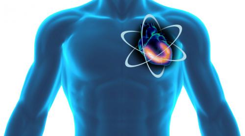
The Essence of Nuclear Cardiologists in Medicine
Nuclear medicine is a branch of clinical medicine that deals with radionuclide pharmaceuticals in diagnostics and treatment. Sometimes, external radiation therapy is also referred to as nuclear medicine. In diagnostics, a cardiologist mainly uses single-photon emission computed tomography (SPECT, captures gamma radiation), positron emission tomography (PET scanners), and radioiodine therapy predominates in the treatment.
The nuclear cardiological diagnosis includes the non-invasive (bloodless) global and regional functional analysis of the heart muscle using the so-called “SPECT myocardial scintigraphy”. With the SPECT myocardial scintigraphy, both the restricted blood flow to the heart muscle in the individual coronary vessels’ supply areas and the global pumping function of the left ventricle can be measured quantitatively.

The purpose of the investigation is based on the two scientific findings that on the one hand, a patient with normal results has an excellent prognosis that cannot be improved by a cardiac catheter examination, a stent, or a bypass operation. On the other hand, a stent or a Bypass surgery only provides a prognostic advantage for a patient if the myocardial scintigraphy shows a circulatory disorder of more than 10% of the heart muscle and, on the other hand, vital heart muscle tissue (myocardium) is still present after a heart attack.
For the examination, a low dose of a weakly radioactive substance (technetium-99m) is injected after dynamic stress (bicycle ergometer) or pharmacological stress. The absorption and distribution of which in the heart muscle is measured using a so-called gamma camera. If a coronary artery is narrowed, there is a regional disruption of isotope uptake, which can then be precisely quantified and topographically assigned to an individual coronary artery.
The diagnostic accuracy of the SPECT myocardial scintigraphy, with a sensitivity of approx. 90%, is significantly higher than the diagnostic accuracy of the stress ECG (sensitivity approx. 50%), which independently of this does not allow an exact topographical assignment to a coronary vessel. The radiation exposure is comparable to computed tomography in X-ray diagnostics and, depending on the examination protocol, is 1 to 4 mSv.
In the nuclear cardiology department of the Heart and Vascular Clinic, the radiation exposure from SPECT myocardial scintigraphy can be reduced to 1 mSv using two new ultra-fast cadmium-zinc-telluride semiconductor detector gamma cameras.
Nuclear cardiology is a form of imaging that uses radioactive indicators and a special camera, called a “gamma camera,” to take pictures of the heart. It provides important diagnostic and prognostic information for the evaluation of patients with heart disease. Combined with a stress test to assess myocardial perfusion, it is instrumental in diagnosing coronary artery disease, establishing its progress, and helping to develop a prognosis. When used, as is often the case, to obtain synchronized images of blood pool activity, it allows the left and right ventricles’ function to be assessed.
Successful performance of nuclear cardiology examinations and careful interpretation of their results requires a good understanding of the principles of nuclear medicine, cardiac physiology, stress testing, and pharmacological stress testing.
Range of services and procedures a nuclear cardiologist takes on a patient
- Preliminary diagnosis of coronary artery disease to clarify the indication for a cardiac catheter examination.
- Evidence of ischemia after a previous cardiac catheterization to clarify the indication for a subsequent balloon dilatation (PCI) / stent implantation or bypass operation in borderline constrictions
- Proof of vitality after a previous cardiac catheter examination to clarify the indication of a subsequent balloon dilatation (PCI) / stent implantation or a bypass operation in the case of extensive infarct scars and reduced left ventricular function
- Follow-up and therapy control after PCI or bypass surgery. After PCI and stent implantation, the dilated vessel can narrow again (restenosis). After a bypass operation, bypass closure occurs in up to 50 percent within ten years—proof of a primarily incomplete revascularizing after PCI or bypass surgery.
- Determination of the global (LVEF%) and the regional left ventricular function as well as the left ventricular volumes (LVEDV, LVESV, and LVSV) with the ECG-triggered gated SPECT myocardial scintigraphy for each myocardial scintigraphy without additional radiation exposure
- Exact quantification of mechanical left ventricular dyssynchrony and regional vitality diagnostics for complete left bundle branch block (LSB) and reduced left ventricular function before possible resynchronization therapy (CRT)
- Detection/exclusion of transthyretin (ATTR) amyloidosis to confirm the diagnosis and possibly plan therapy.
Why is nuclear cardiology essential?
Nuclear cardiology has acquired over the last 20 years an important place in the detection, monitoring, and determination of the prognosis of coronary disease and myocardial viability. The injection of radioactive products, thallium or technetium agents, at rest or after exercise or pharmacological tests makes it possible to study the myocardium’s perfusion. Cardiac tomographic acquisitions are now coupled to the electrocardiogram. They thus make it possible to have the myocardial function simultaneously, that is, to say the ejection fraction of the left ventricle, the regional kinetics, and the volumes of the left ventricle.
The perfusion analysis makes it possible to make the diagnosis of coronary insufficiency with a sensitivity of the order of 90% and a specificity of around 80% in patients with a prevalence of coronary artery disease of 25 to 35%. The importance of perfusion anomalies in size and intensity, the value of the ejection fraction of the left ventricle, the end-diastolic and tele systolic volumes, and regional kinetic disorders make it possible establish a prognosis for the occurrence of coronary events. Thus, a patient who has normal scintigraphy has less than 1% of coronary events per year.
The prognosis is all the worse as the perfusion disorders are essential, and the left ventricular dysfunction is significant. Analysis of the perfusion can also clarify myocardial viability, especially in patients with severe left ventricular dysfunction. It is thus possible to pose any indications for revascularization. A patient has an obvious benefit in survival in the persistence of viability if he is revascularized. This benefit is all the more significant as the ventricular dysfunction is essential. On the other hand, there is no advantage in revascularizing patients without viability. Thus, nuclear cardiology has substantial medical and economic implications in the management of coronary heart patients.


